E. The Vertebral Column

Learning Objectives

  1. Identify each region of the vertebral column and its characteristics.
  2. Compare the general structure of cervical, thoracic, and lumbar vertebrae.
  3. Locate key features using lab skeletons and 3D tools.

Introduction to the Vertebral Column

The vertebral columnβ€”also known as the spinal column or spineβ€”is a flexible, supportive structure that:
🀸 Enables movement🧍 Supports body weight🧠 Protects the spinal cord
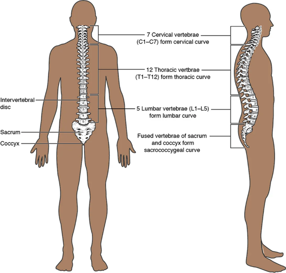
How Many Vertebrae?

The adult vertebral column is typically made up of 33 vertebrae (occasionally 34). These are grouped into five distinct regions:

RegionNumber of VertebraeNotes
Cervical7 (C1–C7)Supports the head, allows rotation
Thoracic12 (T1–T12)Articulates with ribs
Lumbar5 (L1–L5)Largest, bears most weight
Sacral5 (fused)Forms the sacrum
Coccygeal4–5 (fused)Forms the coccyx (tailbone)

πŸ” Note: Only the top 24 vertebrae (cervical, thoracic, and lumbar) are individually moveable. The sacral and coccygeal vertebrae are fused into rigid structures that form the base of the spine.

Use the figure and interactive tools below to gain a spatial understanding of the spine’s regions and curves.

The adult vertebral column (Betts et al., 2022. Figure 7.23 Parts of a Typical Vertebra. In Anatomy and Physiology 2e (2nd ed.). OpenStax. CC BY-NC-SA).

πŸ“Œ What to do:

  • Use the rotation and zoom tools to explore the skeleton from different angles
  • Try identifying each region of the vertebral column in context
  • Observe movement and alignment of the spine in full-body form

πŸ‘‰ Click here to launch the MSD Manual 3D Spinal Column
 Use this tool to visualize vertebral column positioning and see how the spine integrates with the rest of the skeleton.

πŸ‘‰Click here to explore InnerBody’s 3D spinal column. First, select the region of the spinal cord in 2D. Then, select the 3D option to rotate & zoom in to see vertebrae & the spinal cord from multiple angles.

πŸŽ₯ Watch: Overview of the Skeletal System (14:54)

Get a clear, visual introduction to the skeletal system, including the structure and function of bones, joints, and major components like the vertebral column. Ideal for reinforcing lab concepts with 3D visuals and movement demonstrations.

General Vertebral Anatomy


All vertebrae share a common structural plan, though their size and shape vary by region. This section introduces the key features found in most vertebrae and explains their functions. Explore the structure and function of each vertebral region using the interactive tools below

Study the figure below to identify common features on the three types of vertebrae.
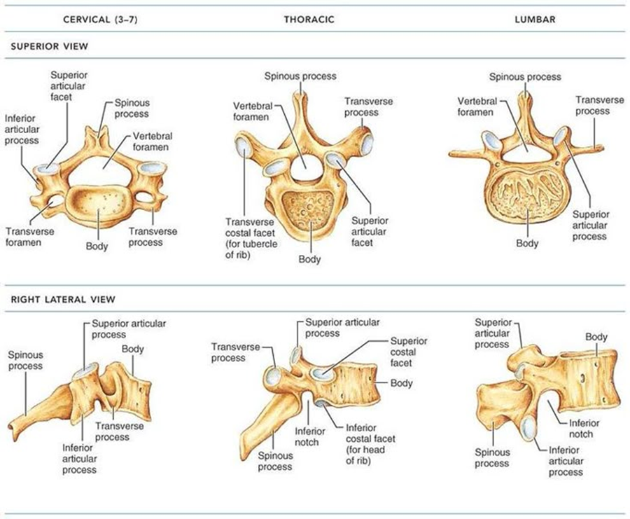
Marieb, E. N. & Hoehn, K. (2007). Human anatomy & physiology (7th ed.). San Francisco: Pearson Benjamin Cummings.

πŸ”Ž Key Vertebral Structures

Vertebral Body

  • The main, load-bearing portion of the vertebra.
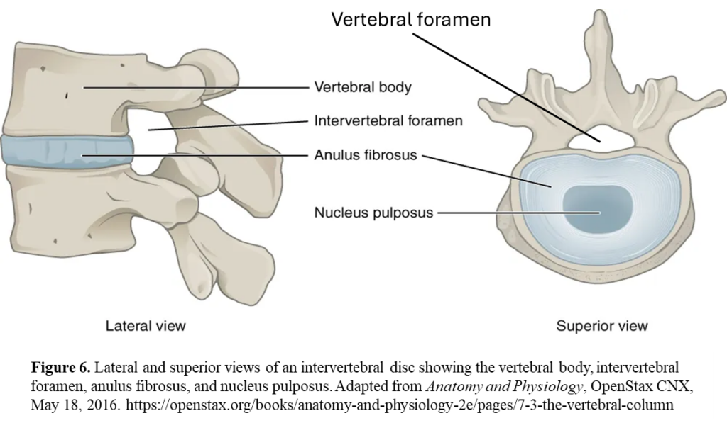
  • Bodies are separated by intervertebral discs, which cushion and allow flexibility.

❓ Question: How does the size of the vertebral body change from cervical to lumbar regions?

Vertebral Foramen

  • The central opening through which the spinal cord passes.
  • Collectively forms the vertebral (spinal) canal for protection and passage of the spinal cord.

Spinous Process

  • Projects posteriorly.
  • In cervical vertebrae, it may be bifid (split tip).

πŸ“Έ Compare bifid vs. single spinous processes using lab models or 3D tools.

Transverse Processes
Extend laterally. In cervical vertebrae, they contain transverse foramina for blood vessels.

Pedicles and Laminae
These form the vertebral arch. Pedicles connect the body to the arch; laminae complete the arch posteriorly. Together, they form the vertebral foramen.

Articular Processes

  • Superior: Point upward; facets face posteriorly.
  • Inferior: Point downward; facets face anteriorly.
    These form synovial joints between vertebrae.

Intervertebral Foramina
Openings between adjacent vertebrae for spinal nerves to exit. Compare these with the vertebral foramen on the figure below.

βœ… Once you’re familiar with these structures, move to the interactive models to test your understanding visually.

It is now time to dive into vertebral details.

πŸ“Œ What to do: Use the descriptions provided in the following five dropdowns as a guide before you explore the 3D features of vertebrae in later exercises.

The vertebral body is the large, anterior portion of each vertebra that bears weight. Notice how the size and shape of the vertebral bodies changes from cervical β†’ thoracic β†’ lumbar.

The higher vertebrae tend to have smaller bodies, and the lower ones (especially lumbar) are larger and more massive to bear weight.

The body of each vertebra is separated from the adjacent ones by a fibrocartilaginous intervertebral disc.

In the 3D view, locate the central opening (foramen) through which the spinal cord passes.

Pay attention to how its shape and relative size may vary across regions.

Transverse processes extend laterally; look for them in all vertebrae.

Spinous processes extend posteriorly; observe differences in length, orientation, and whether they are bifid (split) in cervical vertebrae.

Articular Processes (Superior and Inferior)

Each vertebra has paired superior and inferior articular processes that are bony projections extending from the junction of the pedicle and lamina.

  • Superior articular processes project upwards (superiorly) from the junction of the pedicle and lamina.
  • Inferior articular processes project downwards (inferiorly) from the same junction on the vertebra below.

Alignment with Neighboring Vertebrae:

Each articular process contains a facet (a smooth cartilage-covered surface) that forms synovial joints with the vertebrae above and below. These facets guide and limit spinal movement.

The superior articular facets face posteriorly (and slightly medially in lumbar vertebrae), while the inferior facets face anteriorly.

Cervical vertebrae (C1–C7): small bodies, large foramina in transverse processes (transverse foramina), possibility of bifid spinous processes, relatively delicate structures.

Thoracic vertebrae (T1–T12): medium bodies, long spinous processes that angle downward, facets for rib articulation.

Lumbar vertebrae (L1–L5): large, robust bodies; short, thick spinous processes; no rib facets.

Explore the sacrum and coccyx regions in the model: note how individual vertebrae are fused into a solid structure.

πŸ“Œ Tips:

  • Use this 3D section after reviewing the text-based descriptions and figure above.
  • Rotate, zoom, and isolate each part to reinforce visual-spatial understanding.
  • Ideal for pre-lab review or group study sessions.

1. AnatomyZone: Vertebral Column 3D Explorer

Explore cervical, thoracic, and lumbar vertebrae interactively:

πŸ‘‰ Click here to launch the AnatomyZone Tool.

  • Watch the 7:59 intro video on the vertebral column.
  • Open the 3D rotation tools using the arrow on the bottom right of the image to zoom into different regions.
  • Use the menu on the left and travel through all nine pages.

2. AnatomyTOOL: Compare Vertebrae Across Regions

πŸ‘‰Use this 3D model from AnatomyTOOL to view cervical, thoracic and lumbar vertebrae side by side. This is helpful for comparing:

  • Size and shape of vertebral bodies
  • Spinous process length and angle
  • Presence or absence of transverse foramina
  • Rib facets in thoracic vertebrae

🎯 Goal: Observe and describe 3 major differences between the vertebrae.
(Tip: Jot down your observations to discuss in lab.)

3. AnatomyTOOL: Explore Individual Vertebrae

Use the interactive models below to focus on one vertebral region at a time.

🦴 Cervical Vertebrae 3D β†’ Open model.

🦴 Thoracic Vertebrae 3D β†’Open model.

🦴 Lumbar Vertebrae 3D β†’Open model.

Focus on identifying:

  • Vertebral body
  • Spinous and transverse processes
  • Articular processes (superior/inferior)
  • Vertebral foramen
  • Transverse foramina (in cervical vertebrae)
  • Rib articulation points (in thoracic vertebrae)

Test Your Knowledge

Interactive tools to test your identification skills:

  • Explore 3D images of spinal cord and individual vertebrae
  • Explore Real Bone: View high-resolution images of vertebrae from the TRU lab collection.
  • H5P hotspot activities to label vertebral features.

Click here to explore the spinal column and scroll through all vertebrae. Take note of all regional differences and similarities in vertebral features before completing the exercises below.

Test your identification skills before clicking on the numbers on each vertebra to locate the following:     a) vertebral body  b) vertebral foramen  c) spinous process d) superior articular process e)  inferior articular process f)  intervertebral foramen and g) transverse process

Identify regional features on superior and lateral views of cervical, thoracic and lumbar vertebrae. Then, click on the hotspot to test your recall.