
A. Divisions of the Human Skeleton
Learning Objective: Differentiate between the axial and appendicular skeletons and identify their major components using diagrams, skeletal models, and digital tools.
The skeleton functions to protect some body regions and anchor muscles, making coordinated movements possible. The usual 206 bones are grouped into the following two divisions:
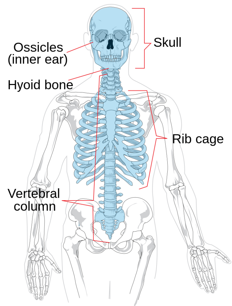
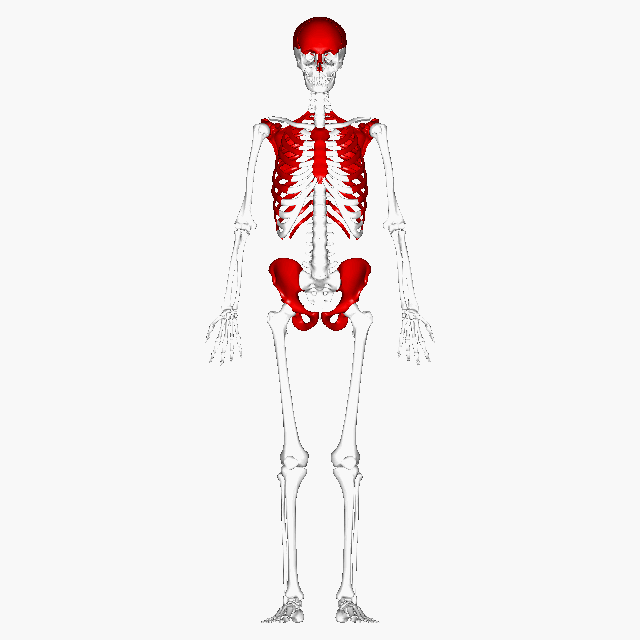
- The axial skeleton consists of the bones of the skull, vertebral column and thoracic cage (Figure 1a). You will learn these first.
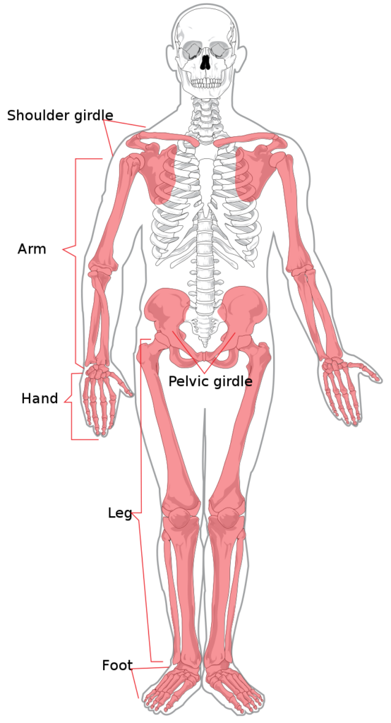
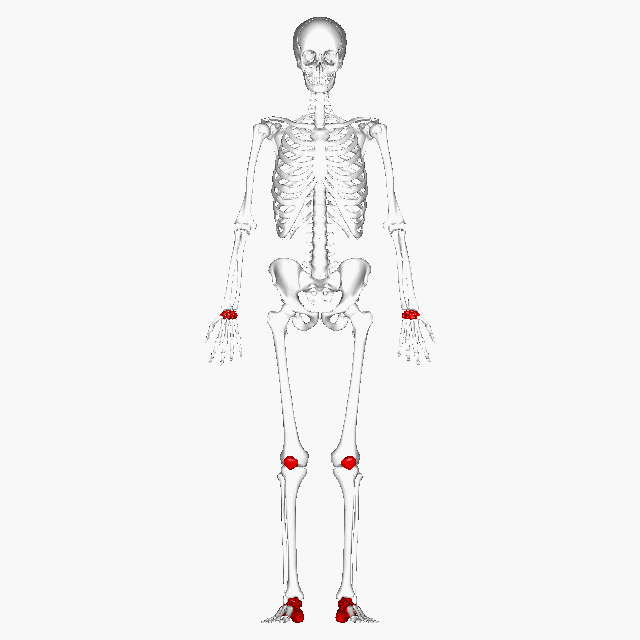
2. The appendicular skeleton is comprised of the bones of the arms and legs, and the two pelvic and shoulder girdles which anchor them to the axial skeleton (Figure 1b).
B. Bone Shapes
Learning Objective: Differentiate between the five bone shape categories and explain how their structures relate to their functions using diagrams, skeletal models, and digital tools.
Betts et al., 2013. A&P 2e OpenStax CC BY-SA 4.0
The 206 bones that compose the adult skeleton are divided into six categories based on their shapes (Figure 2). Their shapes and their functions are related such that each categorical shape of bone has a distinct function.
Bones can be grouped into six shape categories:
- Long bones
- Short bones
- Flat bones
- Irregular bones
- Sesamoid bones
- *Sutural bones
*Note: Sutural bones are small and variable—look closely at cranial sutures in the skull models.
Descriptions
1. Long Bones
A long bone is one that is cylindrical in shape, being longer than it is wide. Keep in mind, however, that the term describes the shape of a bone, not its size. Long bones are found in the arms (humerus, ulna, radius) and legs (femur, tibia, fibula), as well as in the fingers (metacarpals, phalanges) and toes (metatarsals, phalanges). The clavicle is a sigmoid-shaped long bone. Long bones function as levers; they move when muscles contract. Long bones have two main parts; the diaphysis is the central part of the bone and the epiphysis is the rounded end of the bone.
2. Short Bones
A short bone is one that is cube-like in shape, being approximately equal in length, width, and thickness. The only short bones in the human skeleton are in the carpals of the wrists and the tarsals of the ankles. Short bones provide stability and support as well as some limited motion.
3. Flat Bones
The term “flat bone” is somewhat of a misnomer because, although a flat bone is typically thin, it is also often curved. Examples include the cranial (skull) bones, the scapulae (shoulder blades), the sternum (breastbone), and the ribs. Flat bones serve as points of attachment for muscles and often protect internal organs.
4. Irregular Bones
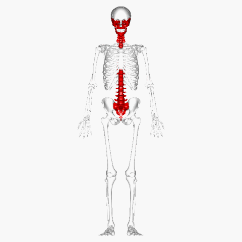
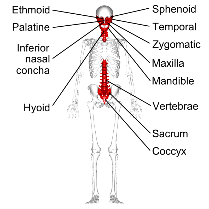
An irregular bone is one that does not have any easily characterized shape and therefore does not fit any other classification. These bones tend to have more complex shapes, like the vertebrae that support the spinal cord and protect it from compressive forces. Many facial bones, particularly the ones containing sinuses, are classified as irregular bones.
5. Sesamoid Bones

/wiki/File:Zohlenanimation.gife
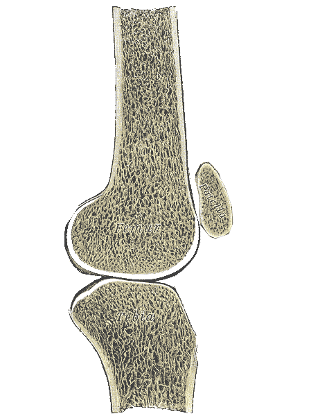
A sesamoid bone is a small, round bone that, as the name suggests, is shaped like a sesame seed. These bones form in tendons (the sheaths of tissue that connect bones to muscles) where a great deal of pressure is generated in a joint. The sesamoid bones protect tendons by helping them overcome compressive forces. Sesamoid bones vary in number and placement from person to person but are typically found in tendons associated with the feet, hands, and knees. The patellae (singular = patella) are the only sesamoid bones found in common with every person.
6. Sutural Bones
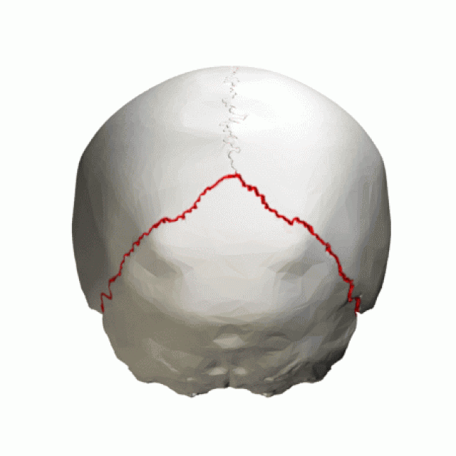
Sutural bones (also called Wormian bones) are small, flat, irregularly shaped bones that form within the sutures (joints) between certain cranial bones. These bones vary in number and size from person to person and are most commonly found in the lambdoid suture at the back of the skull. Sutural bones are not present in all individuals and are considered accessory bones. While their exact function is not fully understood, they may contribute to cranial flexibility during development and help fill gaps between larger bones of the skull.
Table 1 reviews bone shape categories with their associated features, functions, and examples.
Table 1. Bone Shapes Summary
| Bone Type | Shape Features | Function | Examples |
| Long Bones | Longer than wide; shaft and two ends | Leverage for movement | Femur, humerus, phalanges |
| Short Bones | Nearly equal length, width, and thickness | Stability, limited motion | Carpals, tarsals (e.g. cuneiforms) |
| Flat Bones | Thin, flattened, often curved | Protection, muscle attachment | Skull, sternum, ribs, scapulae |
| Irregular Bones | Complex, varied shapes | Protection, support | Vertebrae, facial bones |
| Sesamoid Bones | Small, round, within tendons | Protect tendons, reduce friction | Patella (kneecap) |
| *Sutural Bones | Small, flat, oddly shaped; within sutures of skull | Fill gaps between cranial bones, may aid in skull flexibility | Sutures of the skull (e.g. lambdoid suture) |
*Note: Sutural bones are small and variable—look closely at cranial sutures in the skull models.
Test Your Knowledge
Attributions
Access for free at https://openstax.org/books/anatomy-and-physiology/pages/1-introduction https://en.wikipedia.org/wiki/Appendicular_skeleton https://en.wikipedia.org/wiki/Axial_skeleton
https://en.wikipedia.org/wiki/Long_bone
Gifs By Anatomography is provided by DBCLS – en:Anatomography (setting page of this image at Anatomography), CC BY-SA 2.1 jp, https://commons.wikimedia.org/w/index.php?curid=37817255
“Lambdoid suture – animation05.gif” by Anatomography is licensed under CC BY-SA 2.1 JP.
cup Photo by Danielle MacInnes on Unsplash